
15 Best Radiology Books
The best radiology books, ranked by how many expert sources recommend each one, with notes for medical students, residents, and focused imaging study.
Our take
Radiology books serve different stages of learning: recognizing basic appearances, understanding how images are made, building a systematic search, and comparing possible diagnoses. A medical student usually needs a clear introduction before a large specialty reference. Residents may combine one broad text with books suited to their current rotation. The right choice depends on whether you need explanations, cases, or a practical framework for looking through a study.
Learning Radiology introduces common findings, while Core Radiology offers concise coverage across the specialty. Brant and Helms' Fundamentals of Diagnostic Radiology provides a broader foundation for sustained study. For focused work, Felson's Principles of Chest Roentgenology teaches through questions, Fundamentals of Body CT concentrates on one modality, and Osborn's Brain connects neuroimaging with pathology. Search Pattern and Top 3 Differentials in Radiology address different tasks: looking systematically and comparing diagnoses. Start with an introductory or general text that matches your training, then choose a focused companion.
15 books · 8 sources · Last updated
As an Amazon Associate, we earn from qualifying purchases (at no extra cost to you).
How to choose from this list
Skip to the full rankingIf you're starting with basic interpretation: Learning Radiology
If you want a broad residency foundation: Core Radiology → Brant and Helms' Fundamentals of Diagnostic Radiology
If you're learning chest radiographs: The Chest X-Ray → Felson's Principles of Chest Roentgenology
If you're beginning body CT: Fundamentals of Body CT
If you want to improve your review process: Search Pattern → Top 3 Differentials in Radiology
If you want imaging physics followed by revision: The Essential Physics of Medical Imaging → Review of Radiologic Physics
The 15 books, ranked
Ranked by how many expert sources name each book. Ties go to the book more people recommend.

Brant and Helms' Fundamentals of Diagnostic Radiology
For residents who want a substantial foundation spanning the major imaging specialties. The broad explanations support systematic study beyond isolated cases, though the size makes it less convenient for rapid revision than a concise visual review.
What it's about
This diagnostic radiology reference surveys major imaging specialties, including neuroradiology, chest, breast, abdominal, musculoskeletal, pediatric, and nuclear imaging. Illustrations and review material connect imaging methods with clinical applications, supporting study and reference across a broad range of radiological practice.

For medical students and new trainees who need a first framework for looking at images. Herring emphasizes recognizable findings and systematic comparison, though the introductory scope will not replace the depth needed for specialist training.
What it's about
William Herring introduces radiology through pattern recognition and comparison of normal and abnormal images. Chapters explain common imaging modalities, systematic interpretation, characteristic disease findings, and frequently encountered clinical problems, with images and review questions supporting practice.

For residents who prefer images and compact explanations across the specialty. Mandell's visual organization makes it useful for review and orientation, though the abbreviated coverage often needs supplementing with a fuller reference for unfamiliar topics.
What it's about
Jacob Mandell presents a visual guide to diagnostic imaging using clinical images, color illustrations, and concise explanations. Coverage spans the major radiology specialties, interventional techniques, and imaging physics, providing a resource for learning and examination review.

For students and early trainees who want chest imaging taught through active questions rather than uninterrupted reading. The programmed sequence builds recognition step by step, though its focused teaching format offers less breadth than a comprehensive thoracic reference.
What it's about
Lawrence Goodman teaches chest imaging through a sequenced, interactive approach. Radiographs, annotated examples, questions, and quizzes develop recognition of normal anatomy and disease signs, progressing through chest radiography, CT, lung patterns, pleural spaces, and cardiovascular findings.

Fundamentals of Body CT
For trainees beginning body CT who need anatomy, technique, and common findings brought together. The concentrated modality focus provides a manageable foundation, though more complex cases and specialized protocols require a larger or more focused reference.
What it's about
W. Richard Webb and his coauthors introduce the performance and interpretation of body CT. Illustrated chapters cover thoracic, abdominal, pelvic, and musculoskeletal imaging, emphasizing normal anatomy, scanning techniques, common diseases, and findings that distinguish competing diagnoses.
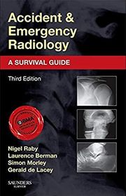
Accident and Emergency Radiology
For clinicians and trainees concentrating on emergency radiographs and injuries. The consistent chapter structure makes anatomy, pitfalls, and overlooked findings easy to revisit, though the practical focus does not offer a complete account of emergency imaging across modalities.
What it's about
Nigel Raby and colleagues present an approach to interpreting emergency radiographs, comparing normal anatomy with injuries and important variants. Chapters use examples, explanatory drawings, and key points to highlight common errors, overlooked abnormalities, and difficulties in diagnosis.
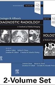
Grainger & Allison's Diagnostic Radiology
For residents who want a comprehensive reference organized around organs and systems. The integrated coverage helps connect imaging methods with clinical applications, though the two-volume scale makes it a longer-term study resource rather than a quick introduction.
What it's about
Andy Adam and fellow editors bring together diagnostic imaging across organ systems and modalities in a two-volume reference. Illustrated chapters connect techniques with clinical applications, including cardiac, functional, and molecular imaging and image-guided procedures, supporting broad radiology training and reference.

Osborn's Brain
For residents who want brain imaging connected closely with anatomy and pathology. Osborn's annotated images support understanding across disease categories, though the detail and specialist vocabulary make this a substantial reference rather than an introductory overview.
What it's about
Anne Osborn and her coauthors connect brain anatomy, pathology, and imaging to explain how neurological diseases appear radiologically. Coverage progresses from trauma, hemorrhage, and stroke to infection, tumors, metabolic disorders, degeneration, and congenital malformations, with annotated images.
Review of Radiologic Physics
For residents who have studied imaging physics and want a concise review of the essentials. Huda keeps attention on image formation, quality, and radiation considerations, though the compressed explanations work best alongside a comprehensive physics textbook.
What it's about
Walter Huda reviews physics relevant to radiological imaging, covering X-rays, fluoroscopy, CT, nuclear medicine, ultrasound, and MRI. The concise text focuses on image creation, image quality, radiation risks, and related quantities, with illustrations and appendices supporting revision.
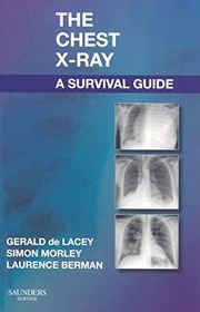
The Chest X-Ray
For medical students and early trainees who want a practical, systematic approach to a common investigation. The emphasis on normal anatomy, errors, and checklists supports focused learning, though its scope is narrower than a general radiology introduction.
What it's about
Gerald de Lacey, Simon Morley, and Laurence Berman explain chest radiograph interpretation using clinical problems, checklists, and annotated examples. Radiographs paired with drawings clarify anatomy and abnormalities, with attention to common mistakes and difficult appearances encountered in practice.

For residents and radiologists who need detailed pulmonary and cardiovascular imaging in one reference. Webb and Higgins connect the findings with disease processes, though the specialist depth is more than most beginners need for learning chest radiographs.
What it's about
W. Richard Webb and Charles Higgins provide a detailed reference for imaging the lungs and cardiovascular system. Illustrated coverage addresses congenital and acquired disease, pulmonary infections, diffuse lung disorders, mediastinal masses, coronary imaging, and myocardial and pericardial conditions.

For trainees who want to practice narrowing an imaging differential through cases. The ranked possibilities make comparisons explicit, though the compressed format is better for checking and consolidating knowledge than learning an unfamiliar subspecialty from scratch.
What it's about
William T. O'Brien presents radiology cases with clinical histories, images, and ranked differential diagnoses. Organized by subspecialty, each case pairs key findings with three leading possibilities, additional considerations, and practical imaging points for clinical and examination review.
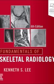
Fundamentals of Skeletal Radiology
For early trainees who want a manageable introduction to musculoskeletal imaging. The sixth edition extends the familiar concise approach across radiographs, MRI, CT, and ultrasound, though detailed subspecialty questions still need a larger skeletal or modality-specific reference.
What it's about
Kenneth Lee's sixth edition introduces skeletal imaging through radiographs, MRI, CT, and ultrasound. Chapters cover trauma, bone tumors and their mimics, arthritis, metabolic conditions, and selected spine and joint examinations, using illustrated examples to connect appearances with interpretation.

For trainees who already know basic findings and want a repeatable way to review studies. Tu's checklists focus attention on search habits and blind spots, though they complement rather than replace explanations of disease and imaging physics.
What it's about
Long H. Tu organizes common radiological studies into structured, step-by-step search patterns. The guide combines reported blind spots with practical expertise to help readers examine images systematically, covering major imaging specialties and workflow checklists for those with basic radiology knowledge.
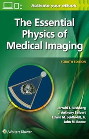
The Essential Physics of Medical Imaging
For residents and medical physics students who need to understand how imaging systems produce their results. The fuller treatment connects physical principles with image quality and radiation protection, though the technical depth requires more time than an exam review.
What it's about
Bushberg and colleagues explain the physical principles underlying medical imaging, including X-rays, CT, MRI, ultrasound, and nuclear medicine. The text connects how images are produced with image quality, radiation interactions, radiation biology, and protection, supported by diagrams and examples.
Sources
- 1
Recommended Radiology Books (2026)
radiogyan.com
- 2
Top Radiology Books — The Radiology Review
theradiologyreview.com
- 3
Radiology ST1 starters guide - Radiology Cafe
radiologycafe.com
- 4
General - Radiology - Specialty Guides at NYU Health Sciences Library
hslguides.med.nyu.edu
- 5
Radiology Books
goodreads.com
- 6
Just started Radiology residency- what books worth it to buy? : r/Residency
reddit.com
- 7
Radiology - Health Sciences Textbooks - Library Guides at University of Missouri Libraries
libraryguides.missouri.edu
- 8
Books - Radiology - Guides at Moody Medical Library, The University of Texas Medical Branch
guides.utmb.edu







Edited by
Software engineer whose passion for tracking book recommendations from podcasts inspired the creation of MRB.
Lead investor at 3one4 Capital whose startup expertise and love for books helped shape MRB and its growth.
Comments
Spotted a missing book, a wrong order or a mistake? Tell us and we'll fix this page.
We're a reading guide, not the author or publisher, so we can't send books or pass on messages.