
15 Best Anatomy Books
The best anatomy books, ranked by how many expert sources recommend each one, with notes for beginners, medical students, and learners choosing a study tool.
Our take
Anatomy books explain the structures of the body and how those structures relate to one another. An illustrated atlas helps you trace a nerve or identify a muscle, while dissection photographs show how tissues look in a specimen. Explanatory textbooks add clinical context, and coloring books offer a more active way to study names and relationships. These formats usually complement one another.
Gray’s Anatomy for Students and Clinically Oriented Anatomy provide explanations to guide a medical course. Netter’s Atlas of Human Anatomy and Gilroy’s Atlas of Anatomy emphasize illustrated reference, while Anatomy by Rohen and colleagues uses dissection photographs. Learners wanting a broader introduction can choose Human Anatomy & Physiology or Anatomy & Physiology For Dummies. For active study, The Anatomy Coloring Book offers a different approach from simply reviewing plates. Start with the depth your course requires, then choose a visual companion that helps you recognize the same structures in another form.
15 books · 9 sources · Last updated
As an Amazon Associate, we earn from qualifying purchases (at no extra cost to you).
How to choose from this list
Skip to the full rankingIf you are new to the subject: Anatomy & Physiology For Dummies
If you are beginning medical study: Gray’s Anatomy for Students → Atlas of Human Anatomy
If you want applied clinical detail: Clinically Oriented Anatomy → Clinical Anatomy
If you are preparing for dissection: Grant’s Atlas of Anatomy → Grant’s Dissector
If coloring helps you learn: The Anatomy Coloring Book
If you study nursing or allied health: Ross and Wilson Anatomy and Physiology in Health and Illness
The 15 books, ranked
Ranked by how many expert sources name each book. Ties go to the book more people recommend.

Atlas of Human Anatomy
For medical students who want a consistent illustrated reference beside a regional course. The clear plates make relationships easier to trace than in photographs, though idealized drawings cannot replace learning to recognize variation in real specimens.
What it's about
Frank Netter's seventh edition presents detailed anatomical illustrations organized to support a clinical understanding of the body. This professional edition connects structures and their relationships through physician created artwork, offering a visual reference for medical and health professions study.

For medical students learning a region for the first time. Its explanations and clinical examples give the pictures a teaching framework, though students preparing for detailed practical identification may still want a separate photographic atlas.
What it's about
Richard Drake, A. Wayne Vogl, and Adam Mitchell organize essential human anatomy by body region in this fourth edition. Illustrations, surface anatomy, diagnostic images, and clinical cases connect structural knowledge with its practical applications in medical study.

For students preparing to recognize structures in the dissection laboratory. Photographs preserve real tissue relationships that drawings can simplify, though they are less immediately clear than an illustrated atlas when you first encounter a region.
What it's about
Johannes W. Rohen, Chihiro Yokochi, and Elke Lütjen-Drecoll's eighth edition presents human anatomy through photographs of cadaver dissections. Regional chapters combine these images with schematics, diagnostic imaging, and clinical cases to explain structures and their spatial relationships.

The Anatomy Coloring Book
For learners who retain names and structures through active visual work. Its coordinated coloring method makes study more participatory than reading an atlas, though it is a supplement and requires time that passive reference does not.
What it's about
Wynn Kapit and Lawrence Elson's fourth edition teaches anatomical structures through coloring activities linked to terminology. By coordinating names with illustrated body parts, the book encourages visual associations and active study rather than relying solely on reading descriptions.

Atlas of Anatomy
For students who want detailed drawings accompanied by tables and explanatory context. Its study-oriented presentation helps connect structures with practical questions, though the volume of material can be demanding if you only need a quick visual lookup.
What it's about
Anne Gilroy and colleagues' fourth edition presents human anatomy through detailed illustrations, radiological images, and explanatory tables. Its organization supports regional study and dissection, with expanded coverage of challenging areas such as the peritoneal cavity and head and neck.

For medical students and junior doctors who want anatomy connected directly with examination and clinical work. Its applied emphasis gives structures a practical purpose, though it offers a different level of visual detail from a dedicated atlas.
What it's about
Harold Ellis and Vishy Mahadevan connect anatomical knowledge with clinical work for medical students and junior doctors. This fourteenth edition uses medical images, diagrams, and illustrated surface anatomy to explain body regions, with expanded coverage of the nervous system.

Clinically Oriented Anatomy
For medical students who want regional anatomy linked closely with clinical situations. Its explanatory approach gives more context than a plate collection, though the extensive detail can make it slow reading alongside a demanding course.
What it's about
Keith Moore, Arthur Dalley, and Anne Agur connect regional anatomy with clinical examination, imaging, and practice in this eighth edition. Illustrations and applied explanations guide students from foundational structures toward understanding their significance in clinical situations.
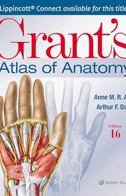
Grant’s Atlas of Anatomy
For students who want illustrations arranged to support regional dissection. Its progression from superficial to deeper structures connects the atlas with laboratory work, though it overlaps substantially with other illustrated atlases and is usually an alternative rather than another necessity.
What it's about
Anne Agur and Arthur Dalley present anatomical illustrations derived from specimens in a regional, surface-to-deep sequence. Supporting text, tables, and clinical material connect the illustrated structures with their relationships and with learning in the dissection laboratory.
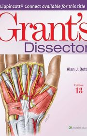
Grant’s Dissector
For students who need guidance through a supervised dissection course. Its sequential instructions serve a different purpose from the finished views in an atlas, though it is a laboratory companion rather than a standalone introduction to anatomy.
What it's about
Alan Detton provides step-by-step instructions for human cadaver dissection, organized by body region. The manual guides preparation, identification, and follow-up study, connecting procedures in the laboratory with the anatomical relationships students are expected to recognize.
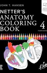
Netter’s Anatomy Coloring Book
For students who like Netter-style illustrations and want to study them actively. Coloring and accompanying explanations help trace structures and relationships, though it overlaps with other coloring books and works best alongside a main course text.
What it's about
John Hansen adapts anatomical illustrations into coloring exercises supported by explanations and review material. Activities guide attention to structures, pathways, and relationships across body systems, offering an active supplement to an anatomy textbook or atlas.

For nursing and health-professions students who need structure, normal function, and illness connected clearly. Its introductory presentation supports broad coursework, though readers seeking the detail of a medical dissection atlas will need another reference.
What it's about
Anne Waugh and Allison Grant introduce human body structure and function, then explain how illness alters normal processes. This twelfth edition uses straightforward text and color illustrations for students preparing for nursing and other health related professions.

For students who need detailed anatomical illustrations for regional study and revision. Its extensive figures and supporting tables offer a substantial reference, though the atlas format assumes you have explanations or teaching to guide your first pass.
What it's about
Reinhard Putz's fourteenth edition of the Sobotta atlas presents macroscopic human anatomy by body region. Detailed figures, introductory overviews, clinical images, and tables of muscles, joints, and nerves connect individual structures with the broader organization of the body.

For students who want a compact regional text with illustrations and self-assessment. Its structured presentation suits review and checking understanding, though the compressed format may feel abrupt when you need longer explanations of unfamiliar material.
What it's about
Anne Gilroy's third edition of Anatomy: An Essential Textbook organizes anatomical knowledge by region, linking illustrations with clinical and developmental correlations. Imaging chapters and extensive questions with explanations support learning, self assessment, and preparation for medical examinations.

Human Anatomy & Physiology
For introductory students who need body structure and function taught together. Its integrated explanations suit a broad course better than an atlas alone, though the physiology coverage adds length for readers interested only in anatomy.
What it's about
Elaine Marieb and Katja Hoehn connect human body structures with their functions in an introductory textbook. Illustrated explanations and clinical applications accompany learning activities designed to help students understand anatomical relationships and physiological processes beyond memorizing terminology.

Anatomy & Physiology For Dummies
For beginners who want unfamiliar terminology explained without the density of a medical textbook. Its conversational approach lowers the entry barrier, though it does not provide the depth or visual detail required for advanced anatomy study.
What it's about
Erin Odya and Maggie A. Norris introduce human body structures and functions through plain language and illustrations. This third edition connects anatomical terminology with explanations of how body systems work, including changes across life and relationships with health.
Sources
- 1
14 Best Anatomy and Physiology Books
bioexplorer.net
- 2
20 Best Anatomy And Physiology Books For Medical Students
themdjourney.com
- 3
Best Resources for Learning Anatomy - Books, Courses & More
instituteofhumananatomy.com
- 4
Anatomy Books
goodreads.com
- 5
ANATOMY BOOK RECOMMENDATIONS PLEASEEEEE
reddit.com
- 6
Vote please, the best Anatomy book for first year students is:
reddit.com
- 7
10 Best Anatomy Books
addall.com
- 8
Anatomy & Musculoskeletal System - Books for COM MSMHS
libguides.tu.edu
- 9
Anatomy: Atlases and Textbooks
guides.library.harvard.edu


Edited by
Software engineer whose passion for tracking book recommendations from podcasts inspired the creation of MRB.
Lead investor at 3one4 Capital whose startup expertise and love for books helped shape MRB and its growth.
Comments
Spotted a missing book, a wrong order or a mistake? Tell us and we'll fix this page.
We're a reading guide, not the author or publisher, so we can't send books or pass on messages.