
15 Best Histology Books
The best histology books, ranked by how many expert sources recommend each one, with notes on who each text or atlas suits, its depth and where to start.
Our take
Histology connects the microscopic appearance of cells and tissues with what they do. Learning it involves both understanding structure and recognizing it in specimens that may look different from an ideal diagram. These books serve medical, dental and biology students, with some extending into the specialist needs of pathology training. A full textbook, an image atlas and a review guide can support different parts of the same course.
Junqueira's Basic Histology and Histology: A Text and Atlas combine explanations with images, while Wheater's Functional Histology keeps tissue appearance and function close together. A Photographic Atlas of Histology offers direct visual comparison with laboratory slides. Histology and Cell Biology connects normal structures with disease, and BRS Cell Biology and Histology is more useful for consolidation and questions. The pathologist’s reference goes deeper into normal variation and assumes an established foundation. Start with Junqueira's Basic Histology for a general course, then use an atlas to practice identifying structures without relying on labels.
15 books · 6 sources · Last updated
As an Amazon Associate, we earn from qualifying purchases (at no extra cost to you).
How to choose from this list
Skip to the full rankingIf you want a general first course: Junqueira's Basic Histology
If you want detailed cellular explanations: Histology: A Text and Atlas
If you want to recognize tissues: Wheater's Functional Histology → A Photographic Atlas of Histology
If you want links with disease: Histology and Cell Biology
If you are reviewing for examinations: BRS Cell Biology and Histology
If you are training in pathology: Mills's Histology for Pathologists
The 15 books, ranked
Ranked by how many expert sources name each book. Ties go to the book more people recommend.

For students who want a detailed course text and atlas in one place. Its integration of microscopic structure with cell and molecular biology supports deeper explanations, though the density can make it slower than a concise laboratory companion.
What it's about
Wojciech Pawlina and Michael Ross combine a histology textbook with illustrated atlas sections. Chapters connect cell, tissue, and organ structure with function and clinical relevance, using labeled photomicrographs and explanations of cellular and molecular biology.

For medical and biology students who want a balanced introduction to tissue structure and function. Its combination of explanations, images and review aids supports course study, though recognizing unfamiliar slides still requires practice beyond reading labeled examples.
What it's about
Anthony Mescher connects human tissue structure and function with cell biology, biochemistry, immunology, and physiology. Light and electron micrographs accompany explanations, summary tables, clinical correlations, and self-test questions in a combined histology text and atlas.

Color Atlas and Text of Histology
For students who want a visual laboratory companion with enough text to orient each image. Its organization makes comparing tissues practical, though the brief explanations are less suited to building a complete conceptual foundation from scratch.
What it's about
Leslie Gartner's atlas combines photographs of microscopic tissues with explanatory text covering major tissue types and body systems. The images and descriptions support laboratory study, helping students identify structures and connect their appearance with organization and function.
Color Atlas of Cytology, Histology, and Microscopic Anatomy
For students who want a compact visual reference across cells, tissues and organs. Its paired images and descriptions make it convenient for quick comparison, though the older presentation and concise text work best alongside current course teaching.
What it's about
Wolfgang Kühnel presents microscopic anatomy through color plates, tissue images and explanatory descriptions. The compact atlas covers cellular structures, tissue types and organ systems, giving medical, dental and biology students a visual aid for recognition and revision.

For students who want normal tissue structure linked closely to disease mechanisms. Its integration with cell biology gives clinical correlations more explanatory weight, though the combined scope can overwhelm readers still learning basic tissue identification.
What it's about
Abraham Kierszenbaum and Laura Tres connect histology and cell biology with pathological abnormalities. Illustrations, diagrams, micrographs, and clinical examples explain normal structures and disease, linking foundational science with concepts encountered in medical study and practice.
Histology and Cell Biology: Examination and Board Review
For students who want targeted self-assessment alongside a histology course. Its focused explanations and questions make review more active than browsing an atlas, though the exam orientation leaves less space for broad conceptual exploration.
What it's about
Douglas Paulsen reviews cell biology, tissue structure and organ systems with clinical connections and practice questions. The sixth edition combines color images with focused explanations and study aids intended to help students identify gaps and prepare for examinations.
Histology: An Essential Textbook
For medical students who want a concise guide built around recognizing structures. Its modular approach supports independent laboratory preparation, though the shorter explanations make it better paired with fuller teaching when cellular mechanisms are unfamiliar.
What it's about
D. J. Lowrie introduces the structure and function of human cells, tissues and organs through concise modules. Slide images, electron micrographs and supporting videos help students connect visual identification with the organization and clinical relevance of normal tissues.
Human Histology
For learners who want to move between whole tissue organization and cellular detail. Its linked low- and high-magnification images make orientation easier, though the compact descriptions provide less conceptual development than a full introductory course text.
What it's about
Bertalan Dudas presents human organs and tissues through full-page photomicrographs linked to higher-magnification views. Compact descriptions identify the structures shown, while the second edition adds organ schematics to support understanding of normal microscopic anatomy.
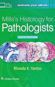
Mills's Histology for Pathologists
For pathology trainees and practitioners who need a detailed account of normal tissues and their variations. Its diagnostic perspective goes beyond undergraduate atlases, though the specialist depth and size make it a poor first histology book.
What it's about
Edited by Rhonda Yantiss, the sixth edition examines normal histology from the perspective of anatomic pathology. Detailed illustrations and organ-based chapters emphasize variations of normal that can cause diagnostic confusion, connecting microscopic structure with its pathological interpretation.

For visual learners who like illustrations connected with micrographs and clinical examples. Its integration of anatomy and tissue detail offers a useful bridge between scales, though stylized drawings need comparison with the variation seen in actual specimens.
What it's about
William Ovalle and Patrick Nahirney connect histology with gross anatomy, embryology, and disease. The textbook and atlas combine classic tissue slides, scanning electron microscopy, illustrations, and histopathology examples to explain microscopic structures and their clinical significance.

For students who primarily need help connecting tissue appearance with function. Its concise explanations and image-centered presentation suit laboratory preparation, though readers wanting extensive molecular mechanisms may need a fuller text alongside the atlas.
What it's about
Barbara Young and her coauthors introduce the microscopic structure of human cells, tissues, and organ systems. Histology images, captions, and concise explanations connect visual features with function, while review questions support study of the fundamentals.

A Photographic Atlas of Histology
For learners who want photographs resembling the ordinary slides used in teaching laboratories. Its visual focus supports recognition practice more directly than lengthy prose, though it provides limited explanation of the molecular and clinical background.
What it's about
Michael J. Leboffe provides a visual reference to human tissues and organs for histology and anatomy study. Photomicrographs of ordinary laboratory slides illustrate structures across the body's systems, helping students compare what they see through the microscope with labeled examples.

Atlas of Histology with Functional Correlations
For students who benefit from comparing idealized drawings with real tissue images. Its functional explanations help translate a diagram into a microscopic view, though the amount of labeling can encourage memorization unless paired with independent slide practice.
What it's about
Victor Eroschenko pairs idealized histologic illustrations with actual photomicrographs to connect tissue appearance and function. The atlas explains cellular and organ structures through functional discussions, chapter summaries, and review questions for medical and allied health students.

For students reviewing after a first pass through cell biology and histology. Its outlines and questions make weak areas easier to identify than a large atlas does, though the compressed format offers less explanation for learning unfamiliar material.
What it's about
Leslie Gartner and Lisa Lee organize cell biology and histology into concise outlines for course and board examination review. Illustrations, tissue images, clinical examples, and practice questions connect basic concepts with their medical applications and provide opportunities for self-assessment.

Textbook of Histology
For students seeking a full narrative course in microscopic anatomy. Its explanations connect tissue organization with cellular mechanisms and clinical relevance, though the comprehensive textbook format is less convenient for quick image checks during laboratory work.
What it's about
Leslie Gartner connects microscopic anatomy with cellular and molecular biology in an illustrated textbook and laboratory guide. Explanations of tissues and organ systems are paired with clinical correlations, showing how histological structure contributes to physiology and disease.
Sources
- 1
72 Best Histology Books of All Time
shortform.com
- 2
MCASOM - Histology: Official Textbooks
libraryguides.mayo.edu
- 3
Books - Histology - University of Queensland Library
guides.library.uq.edu.au
- 4
Books - Pathology & Histology Review - Harvard Library
guides.library.harvard.edu
- 5
Pathologists' Assistant: Histology Resources
libraryguides.quinnipiac.edu
- 6
Histology at SIU
histology.siu.edu




Edited by
Software engineer whose passion for tracking book recommendations from podcasts inspired the creation of MRB.
Lead investor at 3one4 Capital whose startup expertise and love for books helped shape MRB and its growth.
Comments
Did we miss something? Have feedback?
Help us improve this page by sharing your thoughts